
Freshly documented and analysed!
 www.nature.com
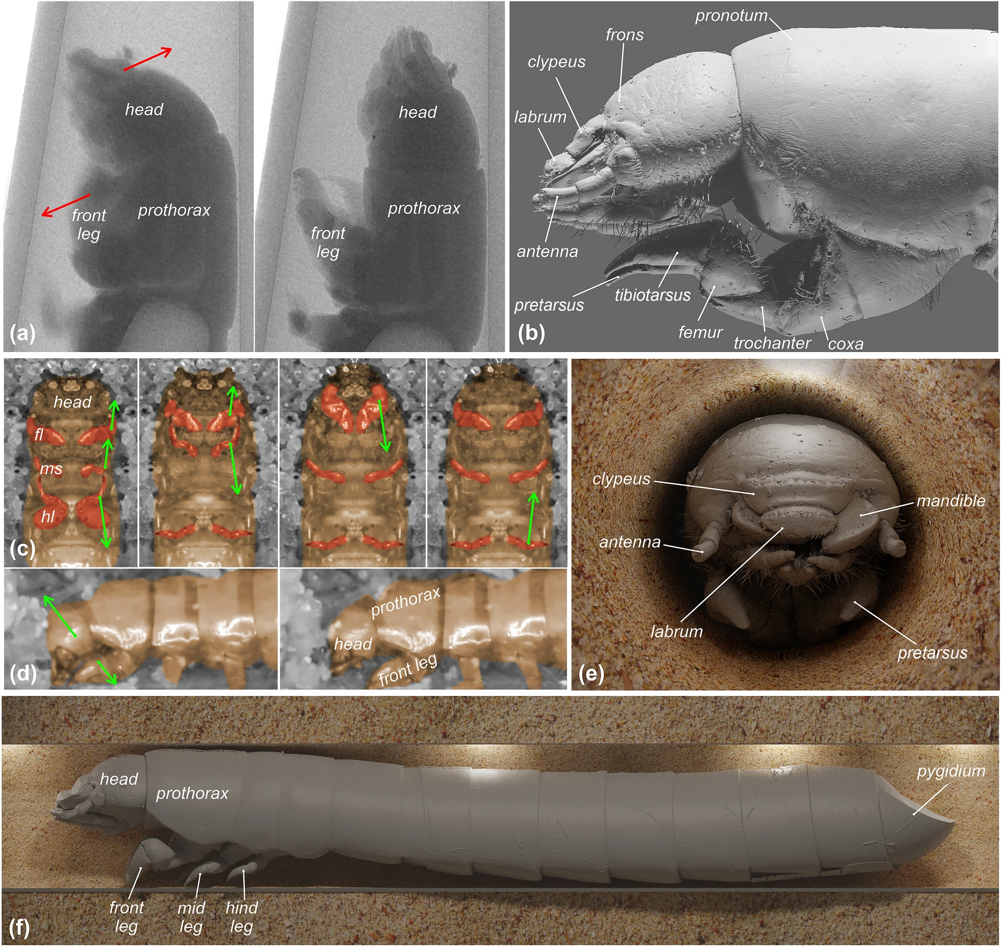
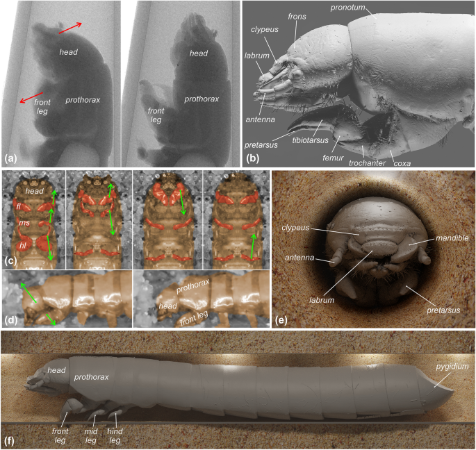
Burrowing dynamics and morphology of the sandworm beetle (Gonopus tibialis) larva. (a) Burrowing images obtained with microCT highlighting changes in head and front legs position. (b) Lateral view of larva imaging the relative position between head and front legs. (c) Ventral and (d) lateral burrowing images (fl—front legs, ml—middle legs, hl—hind legs). (e) frontal and (f) lateral microCT-based visualizations of digging larva. Arrows indicate the motion of particular elements.
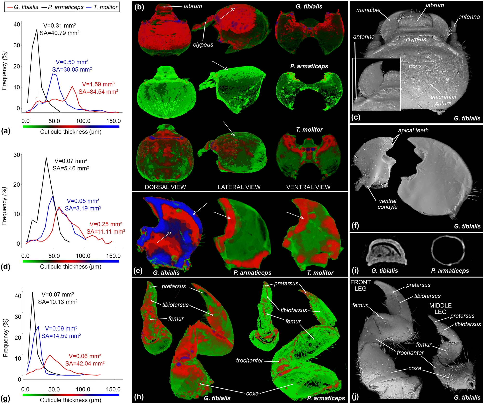
Cuticle thickness analysis dynamics and detailed morphology of larvae of the sandworm beetle (Gonopus tibialis), subsocial beetle (Parastizopus armaticeps), and mealworm (Tenebrio molitor). (a) cuticle thickness distribution of heads, with the exclusion of appendices (i.e. mandibles, antennae, maxillary palpi). (b) MicroCT-based visualizations of cuticle thickness distribution between heads of studied beetle species. (c) head morphology of the sandworm beetle larva. (d) cuticle thickness distribution of rights mandibles. (e) Visualizations of cuticle thickness distribution between right mandibles. (f) mandibular morphology of the sandworm beetle larva. (g) cuticle thickness distribution of front legs (right body side). Arrows indicate major differences between studied species. (h) Visualizations of cuticle thickness distribution between front legs. (i) transverse digital cross sections of front tarsungulus (j) leg morphology of the sandworm beetle larva.
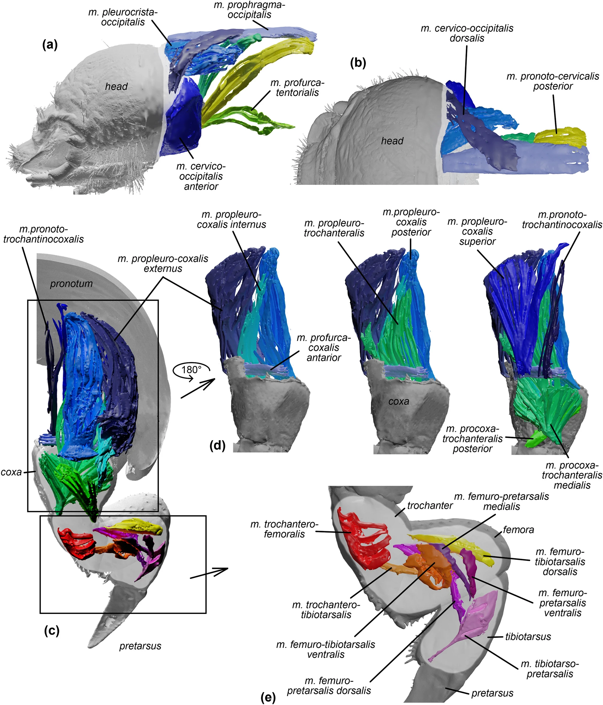
The musculature of the digging apparatus of the sandworm beetle (Gonopus tibialis) larva. (a–c) head: (a) lateral and (b) dorsal views. (c–e) front leg: (c) posterior, (d) frontal, (e) lateral views. Detailed descriptions of particular muscles are presented in Table 1.
Fossoriality in desert-adapted tenebrionid (Coleoptera) larvae - Scientific Reports
In many extreme arid ecosystems, insects constitute major faunal components and are key contributors in nutrient cycling. Previous research on xerophily in insects has focused on adult forms. This study investigates skeletomuscular and behavioural adaptations of the Kalahari sandworm beetle...
www.nature.com In many extreme arid ecosystems, insects constitute major faunal components and are key contributors in nutrient cycling. Previous research on xerophily in insects has focused on adult forms. This study investigates skeletomuscular and behavioural adaptations of the Kalahari sandworm beetle larvae (Gonopus tibialis Fabricius) for dwelling in the sand. Microcomputed tomography enabled cuticle thickness distribution analysis, revealing structural reinforcements of the mandibular edge, the middle part of the head, and the ventral side of the front legs. Laboratory observations and the analysis of muscular system allowed for the definition and functional description of the elements of the digging apparatus of the sandworm larvae. Obtained results point to the crucial role of the head and mandibles in the digging process. These observations are important for understanding desert ecology and pose a challenge to develop newer excavation techniques.
Burrowing dynamics and morphology of the sandworm beetle (Gonopus tibialis) larva. (a) Burrowing images obtained with microCT highlighting changes in head and front legs position. (b) Lateral view of larva imaging the relative position between head and front legs. (c) Ventral and (d) lateral burrowing images (fl—front legs, ml—middle legs, hl—hind legs). (e) frontal and (f) lateral microCT-based visualizations of digging larva. Arrows indicate the motion of particular elements.
Cuticle thickness analysis dynamics and detailed morphology of larvae of the sandworm beetle (Gonopus tibialis), subsocial beetle (Parastizopus armaticeps), and mealworm (Tenebrio molitor). (a) cuticle thickness distribution of heads, with the exclusion of appendices (i.e. mandibles, antennae, maxillary palpi). (b) MicroCT-based visualizations of cuticle thickness distribution between heads of studied beetle species. (c) head morphology of the sandworm beetle larva. (d) cuticle thickness distribution of rights mandibles. (e) Visualizations of cuticle thickness distribution between right mandibles. (f) mandibular morphology of the sandworm beetle larva. (g) cuticle thickness distribution of front legs (right body side). Arrows indicate major differences between studied species. (h) Visualizations of cuticle thickness distribution between front legs. (i) transverse digital cross sections of front tarsungulus (j) leg morphology of the sandworm beetle larva.
The musculature of the digging apparatus of the sandworm beetle (Gonopus tibialis) larva. (a–c) head: (a) lateral and (b) dorsal views. (c–e) front leg: (c) posterior, (d) frontal, (e) lateral views. Detailed descriptions of particular muscles are presented in Table 1.


